This manual facilitates holistic student learning, showcasing innovative features for a diverse experience․ It’s a guide to mastering general biology,
emphasizing safety and practical application within a laboratory setting․
Purpose and Scope
This laboratory manual serves as a comprehensive guide for students undertaking a general biology course․ Its primary purpose is to provide a structured and practical approach to understanding fundamental biological principles․ The scope encompasses a wide range of topics, including microscopy, cell structure, genetics, plant and animal biology, ecology, biochemistry, and microbial studies․
It aims to develop essential laboratory skills, promote scientific inquiry, and foster a deeper appreciation for the living world․ Through hands-on experiments and detailed observations, students will gain practical experience and reinforce theoretical concepts․ This manual supports both traditional and distance-learning environments, adapting to evolving educational needs․
Safety Regulations in the Biology Lab
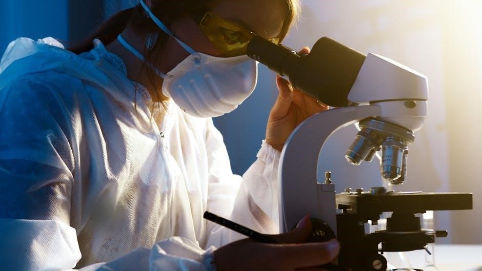
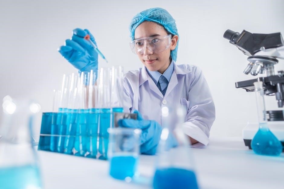
Prioritizing safety is paramount in the biology laboratory․ Always wear appropriate personal protective equipment (PPE), including safety goggles, gloves, and lab coats, to shield against potential hazards․ Never consume food or beverages within the lab space, and refrain from applying cosmetics․
Proper handling and disposal of biological materials, chemicals, and sharps are crucial․ Familiarize yourself with emergency procedures, including the location of safety equipment like eyewash stations and fire extinguishers․ Report any accidents or spills to the instructor immediately․ Maintaining a clean and organized workspace minimizes risks and ensures a safe learning environment for everyone․
Essential Lab Equipment and Their Uses
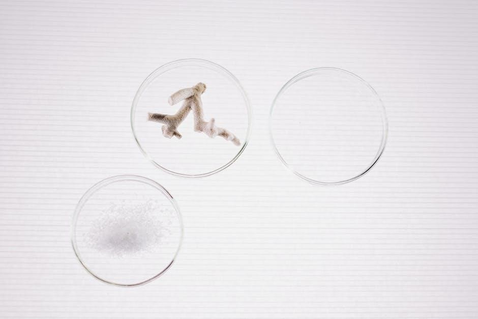
A well-equipped biology lab features diverse tools․ Microscopes – both light and electron – enable cellular observation․ Petri dishes facilitate microbial cultures, while test tubes and beakers are vital for mixing and heating solutions․
Pipettes ensure accurate liquid measurements, and Bunsen burners provide controlled heat․ Scalpels and dissecting tools aid in anatomical studies․ Spectrophotometers analyze light absorption, crucial for biochemical assays․ Understanding the function of each instrument is essential for conducting experiments safely and effectively, yielding reliable results․

Microscopy
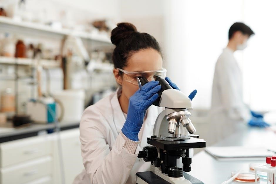
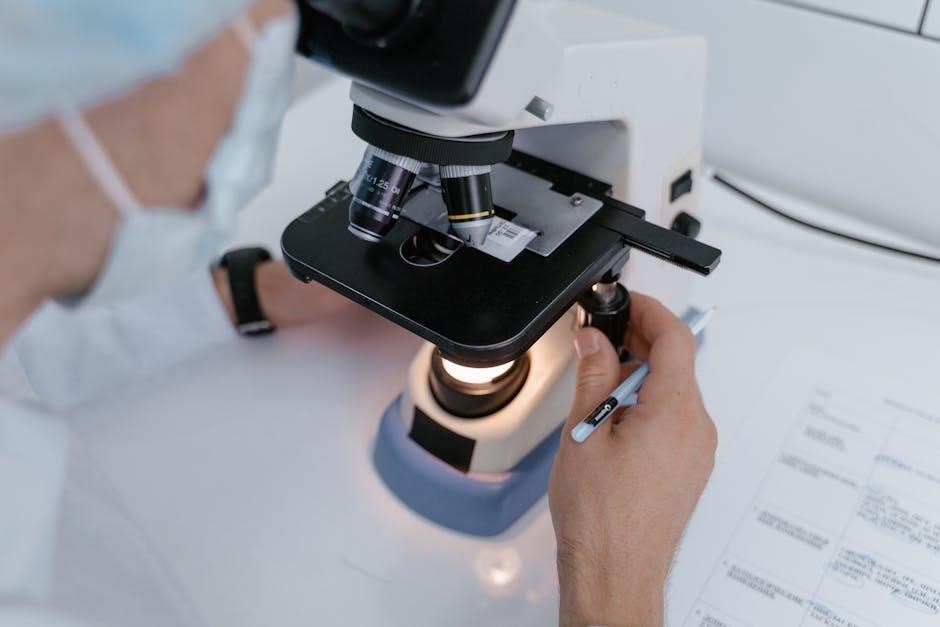
Microscopy unveils the unseen world, utilizing light and electron microscopes for detailed observation of cellular structures and microbial organisms within biological samples․
Types of Microscopes (Light, Electron)
This section details the fundamental types of microscopes used in biological studies․ Light microscopes, employing visible light and lenses, are ideal for observing cells and tissues, offering affordability and ease of use․ However, their resolution is limited․
Electron microscopes, utilizing beams of electrons, provide significantly higher magnification and resolution, revealing ultrastructural details like organelles․ Transmission Electron Microscopy (TEM) examines internal structures, while Scanning Electron Microscopy (SEM) visualizes surface features․
Understanding the principles and applications of each type is crucial for selecting the appropriate tool for specific biological investigations, enabling detailed analysis at varying scales․
Preparing Wet Mount Slides
Creating wet mount slides is a foundational microscopy technique․ Begin with a clean microscope slide and a coverslip․ Place a small sample onto the slide, then add a drop of liquid (water or stain)․ Gently lower the coverslip at an angle to avoid air bubbles, which obstruct viewing․
Excess liquid can be removed with absorbent paper․ Proper wet mount preparation ensures optimal specimen visibility and prevents damage to the microscope objective․
This simple method allows for the temporary observation of living cells and dynamic processes, crucial for introductory biology labs․
Observing Plant and Animal Cells
Microscopic observation reveals the fundamental differences between plant and animal cells․ Plant cells, distinguished by their rigid cell walls and chloroplasts, exhibit a defined shape․ Animal cells, lacking these structures, are more flexible and irregular․
Using prepared slides or wet mounts, students can identify key organelles like the nucleus, cytoplasm, and cell membrane․ Observing these structures provides a visual understanding of cellular function․
Careful examination highlights the structural adaptations that enable each cell type to perform its specific role within the organism․
Cell Structure and Function
This section explores the intricate world of cells, detailing organelle identification and function․ Experiments demonstrate diffusion, osmosis, and enzyme activity, revealing biological processes․
Identifying Cell Organelles
This exercise focuses on recognizing key cellular components through microscopic observation․ Students will examine prepared slides of plant and animal cells, identifying structures like the nucleus, mitochondria, ribosomes, endoplasmic reticulum, and Golgi apparatus․ Detailed diagrams and descriptions will aid in accurate identification․
Understanding the function of each organelle is crucial; therefore, the lab includes questions prompting students to correlate structure with its specific role within the cell․ Emphasis will be placed on differentiating between organelles found in plant versus animal cells, such as the presence of chloroplasts and cell walls․ Proper labeling and detailed observations are essential for successful completion․
Diffusion and Osmosis Experiments
These experiments demonstrate the fundamental principles of passive transport․ Students will observe the movement of molecules across semi-permeable membranes, exploring the effects of concentration gradients, temperature, and molecular size on diffusion rates․ Osmosis will be investigated using potato cores or dialysis tubing immersed in solutions of varying solute concentrations․
Data collection will involve measuring changes in mass or volume over time․ Students will analyze results to determine tonicity (hypotonic, hypertonic, isotonic) and relate observations to biological systems, such as water uptake in plant roots․ Careful controls and accurate measurements are vital for valid conclusions․
Enzyme Activity and Factors Affecting It
This section explores enzyme kinetics through practical investigations․ Students will measure the rate of enzyme-catalyzed reactions, often using catalase breaking down hydrogen peroxide, observing oxygen production․ The impact of substrate concentration, temperature, and pH on enzyme activity will be systematically tested․
Experiments will involve graphing data to determine optimal conditions and understand Michaelis-Menten kinetics․ Students will analyze how deviations from optimal conditions lead to denaturation and reduced activity․ Understanding enzyme function is crucial for comprehending metabolic pathways and biological regulation․
Genetics and Heredity
This module investigates Mendelian genetics using Punnett squares, alongside DNA extraction from fruits․ Observing mitosis and meiosis through microscopy reinforces inheritance principles․
Mendelian Genetics and Punnett Squares
This section delves into the foundational principles of Mendelian genetics, exploring concepts like dominant and recessive alleles, genotype, and phenotype․ Students will actively apply these principles through the construction and analysis of Punnett squares․
These diagrams visually represent the possible combinations of alleles during genetic crosses, predicting the probability of offspring inheriting specific traits․
Practical exercises will involve monohybrid and dihybrid crosses, allowing students to calculate genotypic and phenotypic ratios․ This hands-on approach solidifies understanding of inheritance patterns and the laws of segregation and independent assortment, crucial for comprehending heredity․
DNA Extraction from Fruits
This laboratory exercise provides a tangible experience of isolating DNA from readily available fruit samples, such as strawberries or bananas․ Students will learn the principles behind cell lysis, utilizing household materials like detergent and salt to break down cell membranes and precipitate DNA․
The process involves gently macerating the fruit, followed by filtration and the addition of cold alcohol to visualize the extracted DNA as a white, stringy precipitate․ This hands-on activity demonstrates the physical form of genetic material and the basic techniques used in molecular biology research, fostering a deeper understanding of DNA’s structure․
Observing Mitosis and Meiosis
This lab focuses on visualizing the stages of cell division – mitosis and meiosis – using prepared microscope slides of plant or animal cells․ Students will identify key events like chromosome condensation, spindle formation, and cytokinesis in both processes․
Distinguishing between mitosis, resulting in identical daughter cells, and meiosis, producing genetically diverse gametes, is crucial․ Careful observation and accurate identification of stages will reinforce understanding of these fundamental processes essential for growth, repair, and sexual reproduction․ Diagrams and detailed descriptions aid in accurate stage recognition․
Plant Biology
This section explores plant anatomy, photosynthesis, and transpiration․ Labs involve dissecting roots, stems, and leaves, measuring rates, and analyzing factors influencing these vital processes․
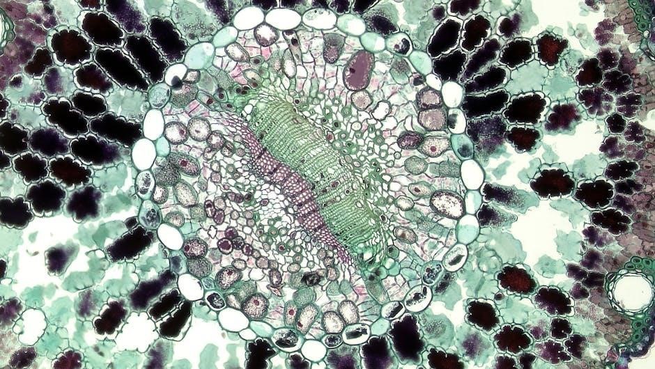
Plant Anatomy: Root, Stem, and Leaf Structures
This lab focuses on the intricate anatomical designs of plants, examining the roles of roots, stems, and leaves․ Students will dissect and observe these structures microscopically, identifying key tissues like xylem, phloem, and parenchyma․
Emphasis is placed on understanding how each structure contributes to the plant’s overall function – absorption, transport, and photosynthesis․
Detailed observations will reveal adaptations related to environmental conditions․
Students will learn to differentiate between monocot and dicot structures, noting variations in vascular bundle arrangement and leaf venation․
Precise labeling and accurate descriptions are crucial for successful completion․
Photosynthesis and Factors Influencing It
This investigation explores the process of photosynthesis, the foundation of most ecosystems․ Students will measure the rate of photosynthesis under varying light intensities and carbon dioxide concentrations, utilizing techniques like floating leaf disk assays․
The lab emphasizes the relationship between light, CO2, and photosynthetic output․
Students will analyze data to determine the limiting factors affecting photosynthetic rates․
Understanding the role of chlorophyll and other pigments is also key․
Detailed observations and accurate data recording are essential for drawing valid conclusions about this vital biological process․
Transpiration Rate Measurement
This lab focuses on transpiration, the process of water movement through a plant and its evaporation from aerial parts․ Students will employ a potometer to quantify transpiration rates in different plant species under controlled environmental conditions․
Variables such as humidity, temperature, and wind speed will be manipulated to observe their impact on water loss․
Data analysis will reveal how these factors influence stomatal opening and closing, regulating transpiration․
Precise measurements and careful observation are crucial for understanding plant water relations and adaptation․
Animal Biology
This section explores animal anatomy through dissection, tissue observation, and physiological process studies․ It provides hands-on experience with representative animal specimens․
Dissection of a Representative Animal (e․g․, Earthworm, Frog)
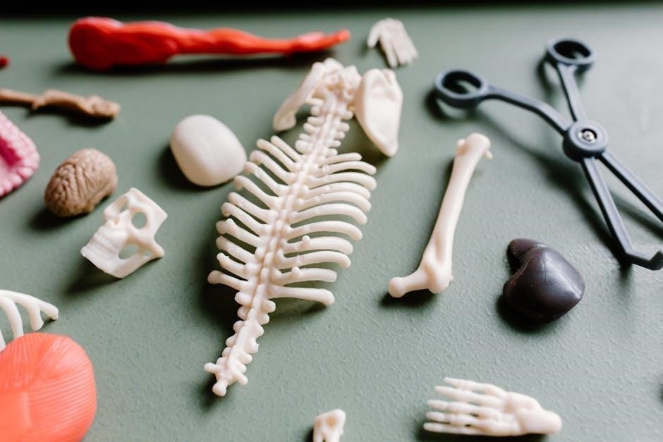
This lab provides a detailed, hands-on exploration of animal anatomy through careful dissection․ Students will systematically examine either an earthworm or a frog, identifying key organ systems and structures․
Emphasis is placed on understanding the relationship between structure and function within the chosen organism․ Proper dissection techniques, safety protocols, and accurate observation skills are paramount․
Detailed diagrams and labeling exercises will accompany the practical work, reinforcing anatomical knowledge․ Students will learn to appreciate the complexity and beauty of animal life through this immersive experience, fostering a deeper understanding of biological principles․
Observing Animal Tissue Types
This laboratory focuses on the microscopic examination of the four primary animal tissue types: epithelial, connective, muscle, and nervous․ Prepared slides will be utilized to observe distinct cellular structures and arrangements characteristic of each tissue․ Students will learn to differentiate between tissue types based on their morphology and function․
Emphasis will be placed on recognizing specialized cells within each tissue and correlating their structure with their specific roles in the body․ Accurate identification and descriptive skills are crucial for success in this lab․
Physiological Processes in Animals
This lab investigates fundamental physiological processes within animals, focusing on key systems like respiration, circulation, and digestion․ Experiments will involve measuring heart rate and blood pressure responses to varying stimuli, simulating digestive enzyme activity, and observing gas exchange mechanisms․ Students will analyze data to understand how these processes maintain homeostasis․
Emphasis will be placed on the interconnectedness of physiological systems and their contribution to overall animal function․ Careful observation and data interpretation are essential for this lab․
Ecology
This section explores ecosystem components, population sampling, and food web construction․ Students will analyze interactions between organisms and their environment, applying ecological principles․
Population Sampling Techniques
Understanding population dynamics is crucial in ecological studies․ This lab introduces various techniques for estimating population sizes within a defined area․ Students will learn and apply methods like quadrat sampling, commonly used for sessile organisms, and mark-recapture techniques, ideal for mobile populations․
These techniques involve collecting data, calculating population density, and analyzing factors influencing population distribution․ Emphasis will be placed on minimizing bias and understanding the limitations of each method․ Practical exercises will allow students to gain hands-on experience in ecological data collection and analysis, fostering a deeper understanding of population ecology principles․
Analyzing Ecosystem Components
Ecosystems are complex, interconnected systems․ This lab focuses on dissecting these interactions by analyzing abiotic and biotic components․ Students will investigate factors like soil composition, water quality, and light intensity – key abiotic elements influencing life․
Biotic analysis includes identifying producers, consumers, and decomposers within a given ecosystem, and assessing their relative abundance․ We’ll explore energy flow through trophic levels and examine the impact of human activities on ecosystem health․ Data collection and interpretation will be central, promoting critical thinking about ecological balance and conservation․
Food Web Construction
Understanding energy transfer is crucial in ecology․ This lab guides students in constructing accurate food webs, illustrating feeding relationships within a specific ecosystem․ We’ll begin by identifying organisms and their trophic levels – producers, primary consumers, secondary consumers, and apex predators․
Students will then diagram these connections, demonstrating the flow of energy and nutrients․ Emphasis will be placed on recognizing the interconnectedness of species and the potential consequences of removing or adding organisms․ Analyzing food web complexity reveals ecosystem stability and resilience․
Biochemistry
Explore life’s molecular basis through experiments testing for proteins, carbohydrates, and lipids․ Investigate pH impacts on biological systems and spectrophotometry’s analytical power․
Testing for Biological Molecules (Proteins, Carbohydrates, Lipids)
This section details qualitative tests to identify key biological macromolecules․ For proteins, the Biuret test utilizes copper ions, producing a violet color in their presence, indicating peptide bond formation․ Carbohydrates are detected using Benedict’s reagent, shifting from blue to green/yellow/orange with increasing sugar concentrations․
Lipids, being hydrophobic, require different approaches; the Sudan III stain dissolves in lipids, creating a red coloration․ These tests provide foundational understanding of biochemical composition, crucial for comprehending life processes․ Careful observation and appropriate controls are essential for accurate results and interpretation within the lab setting․
pH and its Effect on Biological Systems
This experiment explores the critical role of pH in biological functions․ pH, measuring hydrogen ion concentration, profoundly impacts enzyme activity, protein structure, and cellular processes․ Using pH indicators or a pH meter, students will investigate how varying pH levels affect enzyme-catalyzed reactions, observing optimal activity ranges․
The lab demonstrates how deviations from optimal pH can denature proteins and disrupt biological pathways․ Understanding pH’s influence is fundamental to comprehending physiological regulation and maintaining homeostasis within living organisms․ Accurate pH control is vital for reliable experimental outcomes․
Spectrophotometry and its Applications
This lab introduces spectrophotometry, a technique measuring a substance’s light absorption․ Students will learn to utilize a spectrophotometer to quantify the concentration of colored solutions, applying Beer-Lambert Law principles․ The experiment involves creating standard curves and determining unknown sample concentrations․
Applications include analyzing enzyme kinetics, monitoring bacterial growth, and assessing pigment concentrations in plant extracts․ Spectrophotometry provides a quantitative method for studying biological molecules, offering insights into their properties and interactions․ Precise measurements are crucial for accurate data interpretation and analysis․
Microbial Biology
This section details bacterial culturing, identification, and antibiotic sensitivity testing․ Students observe microbial growth, learning techniques vital for understanding these crucial organisms․
Culturing and Identifying Bacteria
This exercise introduces aseptic techniques for cultivating bacteria from various sources․ Students will prepare nutrient agar plates and perform streak plating to obtain isolated colonies․ Morphological characteristics, including colony shape, size, color, and texture, are meticulously observed and recorded․ Gram staining, a crucial differential staining procedure, will be performed to classify bacteria based on cell wall structure․
Further identification relies on biochemical tests, assessing metabolic capabilities like sugar fermentation and enzyme production․ These tests aid in differentiating between bacterial species, providing a foundational understanding of microbial diversity and diagnostic microbiology principles․ Proper disposal protocols are emphasized throughout the process․
Antibiotic Sensitivity Testing
This lab explores the effectiveness of different antibiotics against bacterial isolates․ Students will employ the disk diffusion method, inoculating agar plates with bacteria and applying antibiotic-impregnated disks․ Following incubation, zones of inhibition – clear areas around the disks – will be measured, indicating antibiotic susceptibility․
The diameter of these zones correlates with the antibiotic’s potency; larger zones signify greater sensitivity․ Results are interpreted using established clinical guidelines to categorize bacteria as susceptible, intermediate, or resistant․ This exercise highlights the growing concern of antibiotic resistance and its implications for public health, emphasizing responsible antibiotic usage․
Observing Microbial Growth
This lab focuses on cultivating and visually examining microbial growth patterns․ Students will prepare various growth media – nutrient broth and agar – and inoculate them with bacterial cultures․ Observations will be made over time, documenting colony morphology (shape, size, color) on agar plates and turbidity in broth cultures․
Factors influencing growth, such as temperature and nutrient availability, will be investigated․ Microscopic examination will reveal cellular arrangements and structures․ This exercise demonstrates the rapid reproductive capabilities of microorganisms and the importance of sterile techniques in preventing contamination, crucial for accurate results․
Data Analysis and Scientific Reporting
This section details graphing experimental results and statistical analysis basics․ Students will learn to construct clear visuals and write comprehensive lab reports,
following scientific conventions․
Graphing Experimental Results
Effective data visualization is crucial in biology․ This section guides students through creating accurate and informative graphs from experimental data․ We’ll cover selecting appropriate graph types – line graphs for trends, bar graphs for comparisons, and pie charts for proportions․
Emphasis will be placed on correctly labeling axes, including units, and choosing appropriate scales․ Students will learn to identify and address potential sources of error in their data representation․ Understanding how to present data clearly and concisely is a fundamental skill for scientific communication, enabling effective interpretation and dissemination of research findings․
Statistical Analysis Basics
This section introduces fundamental statistical concepts essential for analyzing biological data․ Students will learn about measures of central tendency – mean, median, and mode – and measures of dispersion, like standard deviation․ We’ll explore the importance of statistical significance and hypothesis testing, including the use of t-tests and chi-square tests․
Understanding these basics allows for objective interpretation of experimental results, distinguishing between genuine effects and random variation․ Emphasis will be placed on selecting the appropriate statistical test based on the data type and experimental design, fostering robust scientific conclusions․
Writing a Lab Report
Effective scientific communication is crucial, and this section details the structure of a standard lab report․ Components include a clear introduction with a defined hypothesis, a detailed methods section outlining procedures, and a presentation of results using tables and figures․
Students will learn to interpret their data and discuss its significance in relation to the initial hypothesis, acknowledging potential errors and limitations․ Proper citation and adherence to scientific writing conventions are emphasized, ensuring clarity and reproducibility of research findings․